Gnome Sciences announces
Linear Tissue Arrays
"A Slice of Life"
Connect with us about Stratified Tissue Microarrays: "A Slice of Life"
What is a Linear Tissue Array?
Gnome Sciences’ Linear Tissue Arrays are larger-format tissue arrays designed to maintain full tissue architecture, including multiple tissue layers (such as epithelial, stromal, and adjacent layers) on a single slide. Unlike traditional TMAs, which use small core biopsies and may miss critical spatial or stratified features, Linear Tissue Arrays retain tissue architecture & structural detail for more comprehensive and biologically relevant analysis.
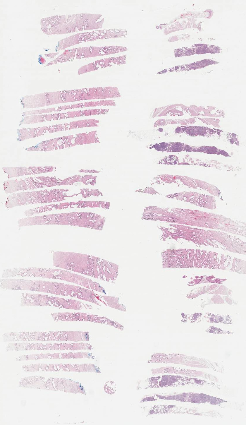
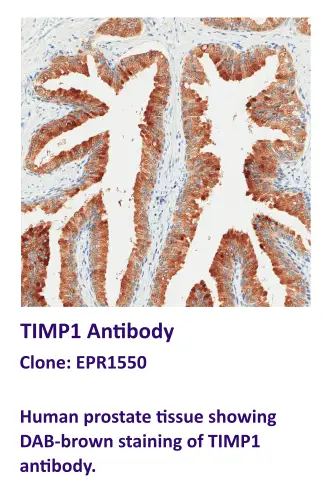
Prostate Tissue Array
To provide a complete picture of tissue architecture, the Linear Tissue Array allows for deeper insights into the function and pathology of tissues with multiple layers.
By maximizing the use of your valuable tissue specimens & antibodies, using the Linear Tissue Array provides a cost-effective solution for large-scale IHC studies & routine diagnostics.
Why it Matters
In many research and clinical contexts, structure tells the story. Whether you are investigating the interface of stratified tissue types (e.g. skin, colon, prostate, etc.), assessing immune infilitration across mucosal layers, or evaluating fibrosis patterns, maintaining spatial & morphological context is essential. Traditional TMAs sacrifice that depth in favor of throughput. Gnome’s Linear Tissue Arrays offer both.
Key Advantages Include:
Preservation of Tissue Architecture & Orientation
Improved Spatial Biomarker Interpretation
Enhanced Reproducibility & Reliability Across Cohorts
Optimized for IHC, ISH, Multiplexing, & Morphology Studies
Applications
Gnome’s Linear Tissue Arrays are ideal for:
- Tissues with multiple layers, such as the skin, colon, prostate, & more
- Biomarker validation across tissue compartments
- Fibrosis, inflammation, & wound healing models
- Drug response studies requiring analysis across multiple tissue layers
How it Works
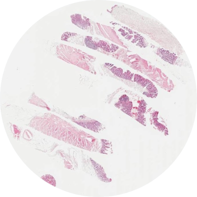
Each array contains full-length tissue sections from multiple donors or experimental groups. Tissue sections are embedded into a single block that can be visualized on a single slide and processed under the same conditions. Arrays can be customized by tissue type, disease state, or experimental endpoint. This format supports highthroughput evalutation with histological precision.
Preserve the Layers. Discover More.
Ready to see the whole picture?

