CISH & FISH
At Gnome Sciences, we provide specialized In-Situ Hybridization (ISH) services to detect and quantify specific DNA and RNA molecules within tissue and cells. Our experienced scientists can perform both Fluorescent and Chromogenic In-Situ Hybridization, providing researchers with the flexibility to select the detection method best suited for their study. From routine target detection to complex multi-marker studies, Gnome Sciences can help design, optimize, and execute ISH assays for a wide range of research applications.
Our dedicated experts are on standby to engage with your project requirements.
Service Overview
Gnome Sciences offers comprehensive In-Situ Hybridization (ISH) services for human and animal tissue, including fresh, frozen, and formalin-fixed paraffin-embedded (FFPE) specimens. Whether you provide prepared slides, tissue blocks, or raw specimens, our scientists can work with you to develop the best approach that accomplishes your study objectives.
Chromogenic In-Situ Hybridization (CISH)
Chromogenic In-Situ Hybridization (CISH) provides a permanent, brightfield-compatible signal of DNA or RNA molecules that can be evaluated using conventional microscopy or digital pathology platforms. CISH can be particularly valuable when nucleic acid detection needs to be integrated with routine histopathology or immunohistochemistry (IHC). Furthermore, CISH uniquely enables spatial visualization of transcript expression across the entirety of an FFPE tissue section. Our scientists can optimize chromogenic detection protocols to provide clear, specific signals while preserving tissue morphology for pathological evaluation.
RNAscope™
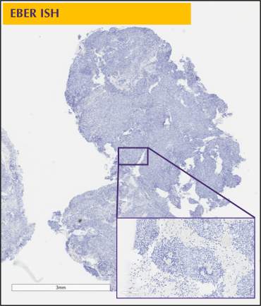
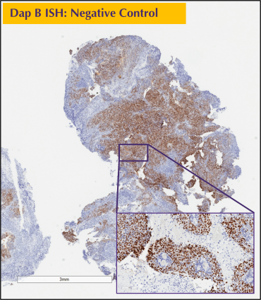
Through collaborations with Advanced Cell Diagnostics (ACD), Gnome Sciences is especially well-versed in performing RNAscope™, a highly specific and sensitive type of CISH. Due to ACD’s patented probe design, RNAscope™ is engineered to drastically amplify the target signal while completely suppressing background noise, enabling for precise signal detection of single RNA transcripts. Gnome Sciences has successfully performed RNAscope™ across a wide array of diverse species and tissue types, including heart, liver, and brain tissue from mice, rats, and non-human primates. As shown here, Gnome Sciences conducted RNAscope™ for the detection of Epstein-Barr virus small RNA (EBER) on nasopharyngeal tissue using DAB (Brown) Chromogen and Hematoxylin (Blue) Nucleus Counterstain. Single EBER RNA molecules can be visualized by the DAB (Brown) signal in the tissue.
Fluorescent In-Situ Hybrdization (FISH)
Our Fluorescent In-Situ Hybridization (FISH) services enable the visualization and localization of specific DNA or RNA sequences within individual cells and tissue sections. Multiple fluorescent labels can be incorporated into a single study to facilitate simultaneous detection and co-localization of targets. Clinical probes can also be utilized with FISH to detect specific genomic alterations and biomarkers associated with clinically relevant disease states. FISH can be applied to a variety of research applications, including gene and genomic analysis, copy number assessment, chromosomal abnormalities, gene rearrangements, and localization of specific nucleic acid sequences.
Multiplex & Co-Localization Studies
Gnome Sciences excels in complex studies requiring simultaneous detection of multiple molecular biomarkers. Multi-color fluorescent approaches can be used to visualize multiple nucleic acid targets within the same section, while chromogenic detection can be combined with immunohistochemistry to evaluate nucleic acid and protein biomarkers together. Our scientists can integrate ISH with other molecular pathology techniques to investigate the spatial relationship between genes, RNA transcripts, proteins, and specific cell populations. For example, Gnome has routinely performed duplex RNAscope™ and neuronal marker IHC staining for the detection and quantitation of single RNA transcripts within the neurons of FFPE brain tissue.
ISH Assay Development & Optimization
Every tissue and target presents unique challenges. Gnome Sciences can assist with probe selection, tissue preparation, assay optimization, signal development, and evaluation of assay specificity and background. We work collaboratively with our clients to identify the most appropriate approach for their research objectives and can optimize existing assays or develop new detection strategies when required.
Digital Imaging & Analysis
ISH studies can be paired with high-resolution digital imaging to document and evaluate molecular signals within tissue. Our automated imaging capabilities allow researchers to visualize target distribution, cellular localization, and co-expression patterns while maintaining the morphological context of the tissue. Depending on study requirements, image analysis from our board-certified pathologists can be incorporated to support qualitative or quantitative assessment of ISH signals. Regardless of your specimen or slide volume, we ensure consistent quality in the staining and scanning of your slides.